Microphthalmia in Small Pets
Microphthalmia (from the Greek mikros, small, and ophthalmos, eye) is a congenital developmental abnormality in which one or both eyeballs are smaller than normal. In the most severe form — anophthalmia — the globe is entirely absent. The condition arises when the embryonic optic vesicle fails to develop normally during fetal life, and the resulting eye may range from mildly reduced in size to a tiny, non-functional remnant buried beneath the eyelids.1
Microphthalmia is not a disease that an animal "catches" or develops over time. It is present from birth, and in the species covered here it is almost always genetic in origin — meaning it is inherited from one or both parents. This has important implications for responsible breeding: animals carrying the relevant mutations should not be used for reproduction, and prospective owners should be aware that certain coat colours and breed types carry a significantly elevated risk.
A small eye is not automatically a painful eye. Many animals with microphthalmia live comfortably with reduced or absent vision in the affected eye, provided the eye surface remains healthy and the animal is not subjected to repeated trauma or infection. The primary veterinary goals are to determine whether the eye is comfortable, to manage any secondary complications (such as corneal exposure or ulceration), and to decide whether the eye should be removed if it becomes a persistent source of pain.
What Causes Microphthalmia?
During normal fetal development, the eye forms from an outgrowth of the developing brain called the optic vesicle. This vesicle invaginates to form the optic cup, which eventually gives rise to the retina, lens, and associated structures. Any disruption to this process — whether from a genetic mutation, a nutritional deficiency during pregnancy (particularly vitamin A), or a toxic or infectious insult during the critical developmental window — can result in a small or absent eye.1
In companion animals, genetic mutations are by far the most common cause. Several specific mutations have been identified in the species discussed below, and in most cases the condition follows a predictable pattern of inheritance that breeders can — and should — act on.
Microphthalmia may occur in isolation, or it may be accompanied by other ocular abnormalities such as congenital cataracts, colobomas (gaps in the iris, retina, or choroid), retinal dysplasia, or malformed eyelids.1 2 The presence of multiple concurrent defects suggests an early and severe disruption of eye development.
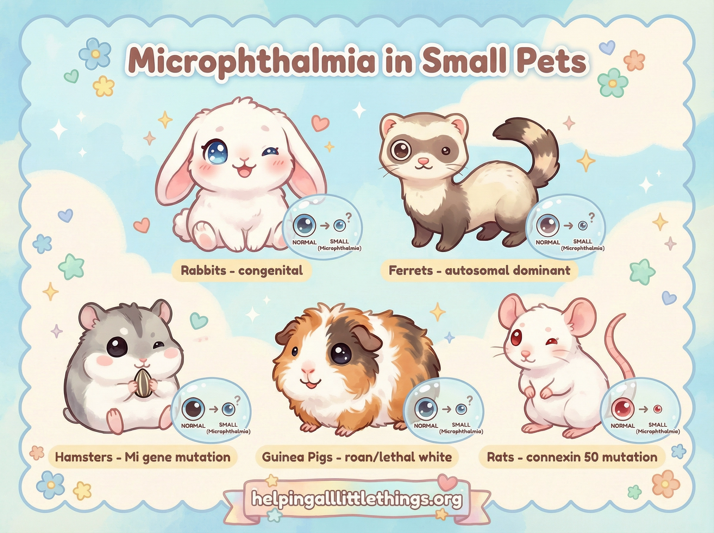
Species-Specific Information
Rabbits
Microphthalmia in rabbits is considered a congenital defect, meaning it is present from birth rather than developing later in life.2 It may affect one or both eyes. The affected eye is visibly smaller than normal and may be accompanied by other structural anomalies, including cataracts, colobomas, or retinal abnormalities. In some rabbits, the small eye retains partial vision; in others, it is entirely non-functional.
A 2025 case report described eyelid agenesis (absent eyelid tissue) in two domestic rabbits from the same litter, with accompanying microphthalmia, iris colobomas, juvenile cataracts, and persistent hyperplastic primary vitreous — illustrating how microphthalmia in rabbits often clusters with other congenital ocular defects.3
Rabbits compensate remarkably well for vision loss, relying heavily on their senses of smell, hearing, and whisker-based spatial awareness. An owner may not notice that a rabbit has a small or non-functional eye until a routine veterinary examination reveals the discrepancy in globe size. The most important clinical questions are whether the eye is comfortable and whether the corneal surface is adequately protected by the eyelids. A microphthalmic eye with poor eyelid coverage is at risk of corneal drying and ulceration, which requires active management.
What to watch for: Persistent squinting, eye discharge, cloudiness of the cornea, or any sign that the eye is causing discomfort. A comfortable, stable microphthalmic eye in a rabbit that is navigating well requires monitoring but not necessarily intervention.
See also: Eye & Vision Issues in Rabbits
Ferrets
Microphthalmia in ferrets is a well-documented autosomal dominant condition, meaning that a ferret needs to inherit only one copy of the mutant gene from one parent to be affected.4 5 The condition was characterised in a laboratory ferret colony and is associated with a syndrome that includes microphthalmos, congenital cataracts, and retinal dysplasia — all arising from the same underlying developmental failure.4
Because the mutation is dominant, it can be passed on by a single affected parent, and it can appear in offspring even when only one parent carries it. This makes the condition relatively easy to propagate inadvertently in breeding programmes that do not screen for it.
Clinically, affected ferrets present with one or both eyes that are visibly smaller than normal, often with a cloudy lens (cataract) visible even in young animals. Vision in the affected eye is typically poor or absent. The condition is not painful in itself, but the associated cataract may progress, and secondary complications such as lens-induced uveitis can develop over time.
What to watch for: Asymmetric eye size noticed at or shortly after eye opening, cloudiness in one or both eyes in a young ferret, or a ferret that navigates poorly in unfamiliar environments. Any ferret with suspected microphthalmia should be examined by a veterinarian experienced with exotic small mammals.
See also: Eye & Vision Issues in Ferrets
Hamsters
Campbell's Dwarf Hamster (Phodopus campbelli)
The most thoroughly documented case of microphthalmia in hamsters involves a specific mutation in the Campbell's dwarf hamster. A 1998 study described the Mi (microphthalmia) gene as an incomplete dominant autosomal mutation.6 The phenotype depends on whether the hamster carries one or two copies of the mutant gene:
| Genotype | Coat Appearance | Eye Appearance | Viability |
|---|---|---|---|
| Wild type (no Mi) | Normal wild-type colouring | Normal eyes | Normal |
| Heterozygote (Mi/+) | Dark markings on white background; slightly lighter than wild type | Black iris with reddish tinge | Normal |
| Homozygote (Mi/Mi) | Pure white all over; smaller body size | Small eyes with unopened eyelids; colourless or faintly reddish iris | Lethal within 3 weeks; rare survivors are sterile |
The homozygous (Mi/Mi) condition is therefore a lethal genotype: affected hamsters also lack incisors and show skeletal abnormalities including dwarfism of individual bones and incomplete fusion of the parietal and frontal skull regions.6 This is directly analogous to the lethal white syndrome seen in guinea pigs (see below).
Heterozygous (Mi/+) Campbell's hamsters are viable and fertile, but their distinctive coat pattern should alert breeders and rescue workers to the presence of the Mi allele in the line. Breeding two heterozygotes together will statistically produce 25% homozygous offspring, all of which will die within three weeks.
Syrian Hamster (Mesocricetus auratus)
Microphthalmia has also been documented in Syrian (golden) hamsters, including a hereditary form associated with colobomatous defects (gaps in ocular structures) and cystic retinal changes.7 Exogenous factors — including irradiation and certain teratogenic agents applied during pregnancy — can also cause microphthalmia in Syrian hamster offspring, making this species a historically important model for studying congenital eye development.8
In practice, a Syrian hamster with a visibly small or absent eye should be assessed by a veterinarian to determine whether the condition is causing discomfort and whether any secondary complications are present.
What to watch for in hamsters: Asymmetric eye size, an eye that does not open fully, persistent discharge from a small or closed eye, or any sign of pain (hunched posture, reduced activity, reluctance to eat).
See also: Eye & Vision Issues in Hamsters
Guinea Pigs
Microphthalmia and anophthalmia (complete absence of the eye) are documented in guinea pigs at a higher frequency than in most other companion species, with the notable exception of inbred laboratory rodents.9 The condition is strongly associated with specific coat genetics.
The Roan/Lethal White Connection
The most important genetic risk factor for microphthalmia in guinea pigs is the roan gene (Rn), which produces the roan coat pattern (a mix of white and coloured hairs). The roan gene is an incomplete dominant: a single copy (Rn/+) produces a roan coat, while two copies (Rn/Rn) produce the so-called "lethal white" phenotype.9
Lethal white guinea pigs are born entirely white (or nearly so) and are characterised by severe congenital defects including microphthalmia or anophthalmia, deafness, and abnormalities of the digestive tract. Most do not survive beyond a few weeks. The condition is directly analogous to the lethal white syndrome in horses and the Mi/Mi genotype in Campbell's hamsters.
Even in heterozygous roan guinea pigs, the prevalence of ocular abnormalities is elevated. A landmark survey of 1,000 guinea pigs found that Abyssinian roan × roan crosses had a statistically significantly higher rate of microphthalmos and cataract than any other breed or cross.9 Eight related animals in one Rex line showed microphthalmos with keratitis, and one individual had clinical anophthalmia with no observable globe structure.
Because of this well-established genetic risk, breeding roan to roan is strongly discouraged by guinea pig welfare organisations. Any roan guinea pig should be paired only with a non-roan partner to avoid producing lethal white offspring.
What to watch for: A visibly small or sunken eye, especially in roan-coloured guinea pigs or their offspring. Secondary keratitis (corneal inflammation) is a common complication when the eyelids do not adequately cover a small globe. Any guinea pig with a small eye should be examined by a veterinarian to assess corneal health and comfort.
See also: Eye & Vision Issues in Guinea Pigs
Rats
Microphthalmia in domestic rats is less commonly reported in the pet context than in laboratory research settings, where it has been studied extensively as a model for human congenital eye disease. However, it does occur in pet rats, and at least one genetic cause has been identified.
A mutation in the connexin 50 gene (Cx50, also known as Gja8) — which encodes a gap junction protein essential for normal lens development — causes autosomal recessive microphthalmia and cataract in rats.10 Affected homozygous rats have significantly smaller eyes and dense cataracts from birth. The connexin 50 protein is critical for the communication between lens fibre cells; without it, the lens fails to develop normally, and the resulting developmental disruption leads to a smaller globe.
In laboratory rat strains, microphthalmia has been documented across fourteen generations of albino rats, suggesting that the condition can persist in a line once established.11 In the pet rat context, the condition is most likely to be encountered in lines that have been selectively bred for unusual coat or eye colours without adequate health screening.
Rats are naturally poor-sighted and rely heavily on their whiskers, smell, and hearing. A rat with microphthalmia in one or both eyes may navigate its familiar environment with little apparent difficulty. The main welfare concern is whether the affected eye is comfortable and whether secondary complications such as corneal ulceration are developing.
What to watch for: Asymmetric eye size, a persistently closed or weeping eye, cloudiness of the cornea, or any sign of ocular discomfort.
See also: Eye & Vision Issues in Rats
Diagnosis
Microphthalmia is typically identified on physical examination when the veterinarian observes that one or both eyes are visibly smaller than normal. A thorough ophthalmic examination will assess:
- Globe size — comparison between the two eyes; measurement with calipers if available.
- Eyelid coverage — whether the eyelids can adequately protect the corneal surface.
- Corneal health — fluorescein staining to detect ulcers or areas of epithelial loss.
- Lens — whether a cataract is present.
- Intraocular pressure — tonometry to rule out concurrent glaucoma.
- Internal structures — ophthalmoscopy or ocular ultrasound to assess the retina and vitreous, particularly if the globe is opaque.
In some cases, referral to a veterinary ophthalmologist is appropriate, particularly if surgery is being considered or if the diagnosis is uncertain.
Management and Quality of Life
There is no treatment that will make a microphthalmic eye grow to normal size. Management is focused on comfort and prevention of secondary complications:
- Lubrication: If the small eye is not adequately covered by the eyelids, artificial tear drops or ointments can prevent corneal drying and ulceration.
- Treatment of secondary infections: Topical antibiotics if bacterial conjunctivitis develops.
- Enucleation (eye removal): If the eye becomes chronically painful, repeatedly ulcerated, or a source of persistent infection, surgical removal is the kindest option. Enucleation eliminates the source of pain and is generally well tolerated by small animals.
- Environmental adjustments: Keep the enclosure layout consistent so the animal can navigate by memory. Avoid sudden rearrangements of furniture, food bowls, and hides.
Most animals with microphthalmia — whether they have partial vision, light perception only, or complete blindness in the affected eye — adapt remarkably well. Prey species in particular are highly reliant on non-visual senses and often show no obvious behavioural signs of visual impairment in a familiar environment.
Breeding Ethics
Because microphthalmia in companion animals is almost always genetic, responsible breeding requires awareness of the relevant risk factors:
- Guinea pigs: Do not breed roan × roan. This cross statistically produces 25% lethal white offspring with severe microphthalmia or anophthalmia, deafness, and digestive abnormalities.
- Campbell's dwarf hamsters: Be aware of the Mi gene in lines with the distinctive white-with-dark-markings coat pattern. Breeding two Mi/+ heterozygotes will produce 25% lethal homozygous offspring.
- Ferrets: The autosomal dominant microphthalmia-cataract-retinal dysplasia syndrome can be transmitted by a single affected parent. Affected ferrets should not be bred.
- All species: Any animal with known microphthalmia should be discussed with a veterinarian before breeding decisions are made.
Rescue organisations should note these genetic risks when assessing animals for adoption and should counsel adopters accordingly.