Entropion in Small Pets
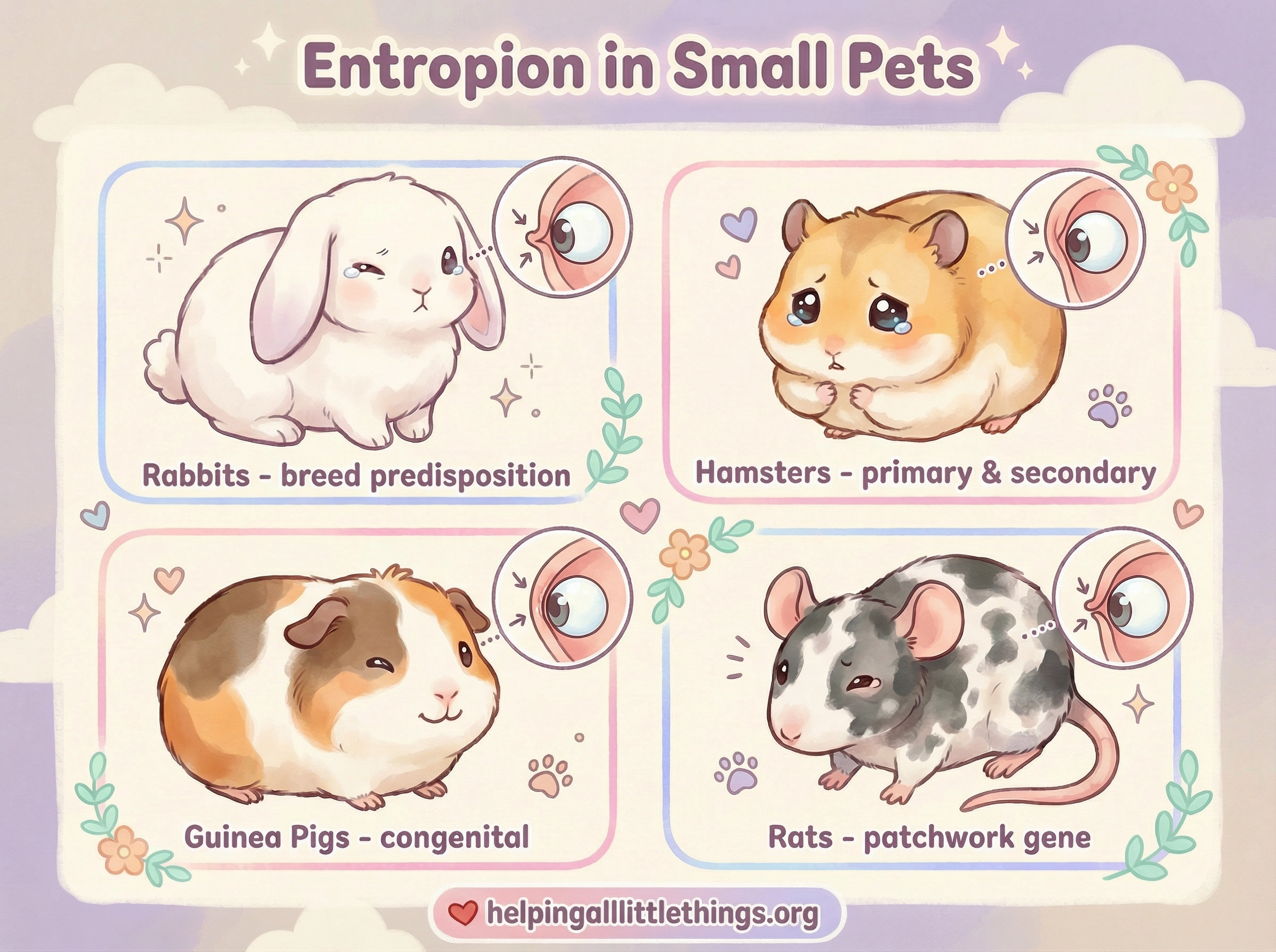
Entropion is a condition in which the eyelid margin rolls inward toward the eye, causing the fur, eyelashes, or skin to rub directly against the cornea (the clear surface of the eye). This constant friction causes pain, irritation, and — if left untreated — progressive corneal damage that can result in scarring, ulceration, and permanent vision loss. Entropion has been documented in several of the species cared for by HALT, including rabbits, hamsters, guinea pigs, and rats.
How Entropion Develops
Entropion is classified according to its underlying cause:
Primary (congenital) entropion is present from birth or develops early in life due to an inherited abnormality in eyelid conformation. The eyelid is structurally predisposed to roll inward. This form is associated with specific breeds in rabbits and with the patchwork (roan/husky) coat gene in domestic rats.
Secondary (cicatricial or spastic) entropion develops as a consequence of another condition. Chronic inflammation of the eyelids (blepharitis), conjunctivitis, or eyelid trauma can cause scarring and contraction of the eyelid tissues, pulling the margin inward. In small rodents, poor husbandry — particularly dusty or irritant bedding — is a recognized contributing factor, as it can trigger the conjunctivitis and blepharoedema (eyelid swelling) that lead to secondary eyelid inversion.1
Species-Specific Information
Rabbits
Entropion is the most commonly observed adnexal (eyelid and surrounding tissue) abnormality in rabbits.2 It can be either primary (congenital) or secondary (cicatricial, developing after chronic blepharitis or other eyelid disease).
Certain breeds have a documented predisposition to primary entropion, suggesting an inherited basis:
| Breed | Eyelid(s) Affected |
|---|---|
| New Zealand White | Lower eyelids |
| French Lop | Upper eyelids |
Secondary entropion in rabbits can develop as a complication of Treponema cuniculi (rabbit syphilis), dacryocystitis (tear duct infection), conjunctivitis, or keratitis.2
Clinical signs include epiphora (excessive tearing), blepharospasm (squinting), conjunctival redness, and — in more advanced cases — corneal ulceration. Applying a topical anesthetic (1% proparacaine) during examination is useful both to allow a thorough assessment and to distinguish true structural entropion from a spastic component that resolves once pain is removed.2
Treatment: Mild cases in rabbits may be managed with topical lubricants and treatment of any underlying infection. Surgical correction using the modified Hotz-Celsus procedure — in which a small crescent of skin is excised from above or below the eyelid margin to evert it — is the standard approach for primary entropion.2 Cicatricial entropion requires a different surgical technique (Y-to-V blepharoplasty) that also removes the fibrotic scar tissue causing the inversion. A minimally invasive alternative — subconjunctival sodium hyaluronate injection — has been reported as a temporary measure in rabbits, providing eyelid eversion without surgery.3
Hamsters
Entropion in hamsters — both Syrian (Mesocricetus auratus) and dwarf species — is recognized in the veterinary ophthalmology literature, though it is less commonly reported than in rabbits.4 Primary entropion has been described in Syrian and dwarf hamsters, and secondary entropion can develop from chronic conjunctivitis, often triggered by dusty or irritant bedding.
A 2025 case report documented the first published surgical correction of entropion in a Syrian hamster, providing important guidance for clinicians.1 The patient was a 3-month-old male hamster presenting with a 3–4 week history of chronic ocular discharge and eyelid inflammation. Examination revealed moderate-to-severe entropion affecting both the upper and lower eyelids at the medial canthus, with associated trichiasis (hairs rubbing the cornea), conjunctival redness, mucoid discharge, photophobia, and persistent blepharospasm. Fluorescein staining was negative, confirming no corneal ulceration had yet developed.
Medical management (topical ciprofloxacin, diclofenac, and systemic meloxicam, combined with husbandry improvements) provided temporary relief but failed to resolve the structural entropion. Surgical correction using the modified Hotz-Celsus technique was performed under general anesthesia. Small ellipses of skin were excised from the medial aspects of both the upper and lower eyelids and closed with 5-0 Monocryl sutures. All sutures healed spontaneously within one week, and at a one-year follow-up, the hamster had complete resolution of ocular irritation and normal eyelid position.1
Important note on anesthetic risk: Surgery in hamsters carries significantly higher anesthetic risk than in larger species. Anesthesia-related mortality in hamsters has been reported at approximately 3.64%, compared to 0.17% in dogs.1 Any surgical procedure in a hamster should be performed by a veterinarian experienced with exotic small mammals, with careful attention to temperature maintenance and respiratory monitoring.
Conjunctivitis is found in approximately 11% of hamsters under primary veterinary care in the UK,1 and because hamsters are nocturnal, owners may not notice early signs of eye irritation until the condition has progressed. Daily observation during the hamster's active evening hours is recommended.
Guinea Pigs
Entropion has been documented in guinea pigs, though it is less frequently reported than in rabbits or hamsters.5 As in other species, it can be primary or secondary to chronic eyelid inflammation. Guinea pigs are also prone to other eyelid and conjunctival conditions — including dermoids (congenital tissue growths on the eyelid or cornea) and "pea eye" (conjunctival prolapse) — that can cause similar signs of ocular irritation and should be differentiated from entropion by a veterinarian.
Rats
Entropion in domestic rats is most notably associated with the patchwork (roan/husky) coat pattern. This coat type, which produces a distinctive patchy or mottled appearance, is linked to a genetic variant that also predisposes affected rats to entropion. The eyelids curl inward so that the fur or eyelashes rub against the cornea, causing chronic pain, corneal ulceration, and potential vision loss.6 In one survey of patchwork rats in Brisbane, Australia, 100% of patchwork rats examined had entropion.6
Because this is a heritable condition directly tied to the patchwork gene, the responsible breeding community strongly advises against breeding patchwork rats, as doing so perpetuates this painful condition. Rescue organizations and ethical breeders should be aware of this association.
Entropion in non-patchwork rats is rare and, when it occurs, is typically secondary to eyelid trauma or chronic inflammation.4
Recognizing Entropion: Signs to Watch For
Regardless of species, the clinical signs of entropion are similar:
- Persistent or recurrent ocular discharge (clear, mucoid, or purulent)
- Squinting (blepharospasm) or holding the eye partially or fully closed
- Redness of the conjunctiva (the pink tissue around the eye)
- Tearing or wet fur below the eye
- Rubbing at the eye with a paw
- Visible inward rolling of the eyelid margin
- Cloudiness or bluish discoloration of the cornea (indicating ulceration — seek emergency care)
Because these signs overlap with many other eye conditions, a veterinary examination is essential for an accurate diagnosis. Never attempt to treat an eye condition at home without a diagnosis, as the wrong treatment can worsen the condition.
Diagnosis
A veterinarian experienced with exotic animals will examine the eyelids and cornea, typically using magnification and a slit lamp or direct ophthalmoscope. Fluorescein staining — a harmless orange dye that highlights corneal defects — is used to check for corneal ulcers, which are a serious complication requiring urgent treatment. Skull radiographs or CT imaging may be recommended in rabbits to rule out dental disease as a contributing factor.
Treatment
Treatment depends on the severity of the entropion and whether corneal damage has occurred:
Medical management is appropriate for mild cases or as a temporary measure:
- Topical lubricants (artificial tear gels) to protect the cornea
- Topical antibiotics if secondary bacterial infection is present
- Topical anti-inflammatory drops (e.g., diclofenac) to reduce swelling
- Husbandry improvements: switching to low-dust, non-irritant bedding; reducing ammonia buildup with more frequent partial cage cleans
Surgical correction is required for structural entropion that does not resolve with medical management. The modified Hotz-Celsus procedure is the most commonly used technique, involving the excision of a small ellipse of skin to evert the eyelid margin. This procedure has been successfully performed in rabbits and, as of 2025, in Syrian hamsters.1 2 Surgery should be performed by a veterinarian with exotic animal surgical experience.
Corneal ulcers, if present, must be treated concurrently with aggressive topical antibiotic therapy and pain management. An untreated ulcer can lead to perforation of the eye and permanent vision loss.
Prevention
- Choose bedding carefully: Use low-dust, non-aromatic paper or aspen bedding. Avoid cedar, pine, or dusty substrates that can irritate the eyes.
- Maintain cage hygiene: Ammonia from urine is a significant ocular irritant. Partial spot-cleans more frequently are preferable to infrequent deep cleans that allow ammonia to accumulate.
- Support responsible breeding: Avoid supporting breeders of patchwork rats or rabbit breeds with known entropion predispositions unless those breeders are actively working to reduce the incidence of the condition.
- Monitor daily: Observe your pet's eyes during their active periods. Early detection of any discharge, squinting, or redness allows for prompt treatment before corneal damage occurs.