Hamster Polyomavirus (HaPyV)
%20Infographic)
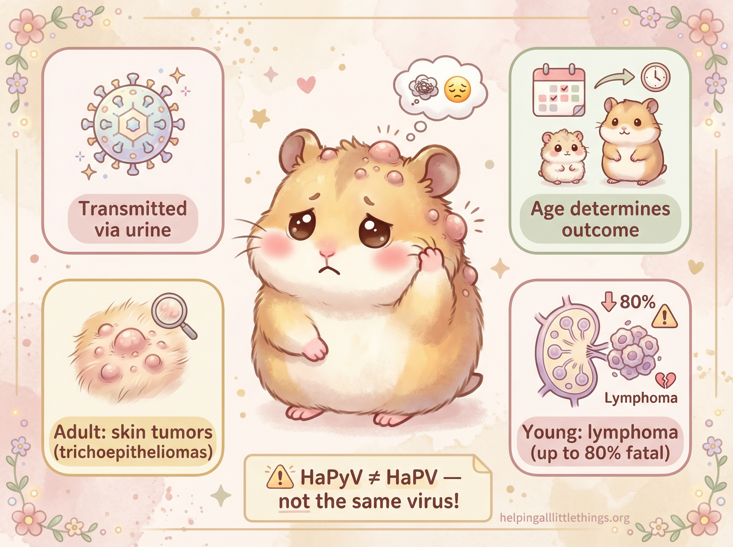
Hamster Polyomavirus — formally known as Mesocricetus auratus polyomavirus 1 and abbreviated HaPyV — is a naturally occurring, highly contagious DNA virus that affects Syrian (golden) hamsters (Mesocricetus auratus). It is the only well-characterized polyomavirus known in hamsters and is the sole cause of a specific type of skin tumor called a trichoepithelioma in this species.1 2 While HaPyV infection is uncommon in laboratory hamster colonies today, it may be encountered more frequently in some pet hamster populations.3
The abbreviation HaPyV (Hamster Polyomavirus) is the current standard and should not be confused with HaPV, which historically referred to both the polyomavirus and the entirely separate Hamster Parvovirus — a completely different virus that causes dental and neurological problems in young hamsters. These two viruses are unrelated. When reading older veterinary literature, always check which virus is being discussed.1
What Is a Polyomavirus?
Polyomaviruses are small, non-enveloped viruses with a circular double-stranded DNA genome. They are classified as oncogenic (tumor-causing) viruses because they carry genes — called tumor antigens (T antigens) — that can drive uncontrolled cell proliferation. HaPyV belongs to the genus Alphapolyomavirus and is phylogenetically related to the murine polyomavirus.1 In humans, related polyomaviruses are responsible for conditions such as Merkel cell carcinoma and progressive multifocal leukoencephalopathy in immunosuppressed individuals, illustrating the clinical significance of this virus family.
How Is It Transmitted?
HaPyV spreads horizontally — that is, between individual hamsters, not from mother to offspring through the egg or placenta. The primary route of transmission is through urine, as the virus persists in the renal tubular epithelium of infected adult hamsters and is continuously shed.2 3 The virus can also spread through biting and grooming behaviors. Environmental contamination is a significant concern: aggressive decontamination efforts in affected research facilities have sometimes failed to eliminate the virus, and increased use of personal protective equipment has also proven insufficient in some outbreaks.1
Because a hamster can shed the virus in urine without showing any outward signs of illness, an apparently healthy animal purchased from a pet store or breeder may already be infected. One documented case involved a hamster assumed to have been infected before purchase at one month of age, as it had been housed alone with no subsequent contact with other hamsters.3
Two Very Different Diseases: Age Determines the Outcome
The most striking feature of HaPyV is that the age of the hamster at the time of infection determines which disease develops. The virus can cause two entirely distinct neoplastic (tumor-forming) syndromes:
| Feature | Trichoepitheliomas (Skin Tumors) | Lymphoma / Leukemia |
|---|---|---|
| Age at infection | Typically older hamsters | Young, immunologically naïve hamsters |
| Tumor type | Benign hair follicle tumors | Malignant lymphoid tumors |
| Affected sites | Skin (head, neck, back, belly, feet) | Mesenteric lymph nodes, liver, spleen, thymus |
| Prognosis | Guarded to poor (debilitating) | Very poor (mortality up to 80%) |
| Spread | Does not metastasize | Invades adjacent tissue; metastasizes |
1. Trichoepitheliomas (Hair Follicle Tumors)
Trichoepitheliomas are the hallmark of HaPyV infection in adult hamsters. HaPyV targets hair follicle keratinocytes — specifically the cells of the hair root epithelium — and drives their uncontrolled proliferation.3 Critically, trichoepitheliomas in hamsters have not been described in association with any cause other than HaPyV infection.2 3 If your hamster develops these tumors, HaPyV is the presumed cause.
What they look like: The tumors begin as firm, alopecic (hairless) nodules, typically first appearing on the head, around the eyes and ears. They then spread to the neck, back, ventral abdomen, limbs, and feet. Over time, multiple nodules can coalesce into massive, confluent thickened layers of skin.1 3 Histologically, the tumors are cyst-like masses filled with cornified (keratinized) material, sometimes containing melanin. The tumors are benign — they do not spread to internal organs — but their progressive size and number can become severely debilitating.3
At any age, infection with HaPyV can be associated with trichoepithelioma development in up to 10% of hamsters, though rates are often higher in colonies where the infection is endemic.3
2. Lymphoma and Leukemia
When young, immunologically naïve hamsters are infected with HaPyV, the outcome is far more severe. The virus infects lymphocytes as well as keratinocytes, driving the development of multicentric lymphoma or leukemia.1 Mortality rates among affected young hamsters can reach 80% within 4 to 30 weeks of infection.1 3
The most commonly affected sites are the mesenteric lymph nodes and liver, with less frequent involvement of the small intestine, thymus, kidneys, spleen, and peripheral lymph nodes.3 Grossly, the masses are typically soft, pale tan, and bulge when sectioned. Histologically, the neoplastic lymphocytes are large and immature; invasion of adjacent tissue and metastasis are common as the disease progresses.3 Both B-cell and T-cell lymphomas have been documented.3
Clinical signs are often nonspecific and referable to the affected organs: weight loss, lethargy, palpable abdominal masses, or respiratory difficulty from large intrathoracic tumors. Because hamsters are adept at hiding illness, these signs may not be apparent until the disease is advanced.
The concurrent development of both skin tumors and lymphoma in the same individual hamster is a rare but documented occurrence.3
Diagnosis
A tentative diagnosis of HaPyV-associated trichoepithelioma can be made based on the characteristic clinical appearance (multiple firm, alopecic nodules beginning on the head) combined with histopathology showing the typical cyst-like, cornified masses.2 Since trichoepitheliomas in hamsters are exclusively caused by HaPyV, the histological diagnosis effectively confirms viral involvement.
For lymphoma, a PCR assay can be used to identify HaPyV DNA in transformed neoplastic lymphocytes.3 Electron microscopy can detect viral particles within trichoepitheliomas. Serological testing (indirect ELISA and Western blot) can detect antibodies against the VP1 capsid protein.1
Treatment and Prognosis
There are currently no treatment options for HaPyV infection itself. The virus cannot be cleared once established.1 Management is therefore focused on:
- Trichoepitheliomas: Individual tumors may be surgically excised to improve quality of life and reduce discomfort, but new tumors will continue to develop as the infection is not eliminated. The progressive nature of the disease means prognosis is guarded.
- Lymphoma: The prognosis is very poor. Supportive care may be offered, but the aggressive nature of HaPyV-associated lymphoma and the high mortality rate mean that euthanasia is often the most humane option once the disease is advanced.
In research colony settings, culling of all animals is the primary recommendation when HaPyV infection is widespread, as environmental decontamination has proven unreliable.1
What Should You Do If You Suspect HaPyV?
If your hamster develops multiple firm, hairless lumps — particularly starting around the face — or shows signs of weight loss and abdominal swelling, consult an exotic animal veterinarian promptly. While the diagnosis may be distressing, early veterinary involvement allows for proper pain management, quality-of-life assessment, and guidance on when humane euthanasia may be appropriate.
If you have multiple hamsters, strict isolation of the affected individual is essential, as the virus spreads readily through urine and contact. Be aware that apparently healthy cagemates may already be infected and shedding the virus.
Prevention
There is no vaccine available for HaPyV. Prevention relies on:
- Sourcing hamsters from reputable breeders who screen their colonies and maintain good biosecurity.
- Quarantining new hamsters before introducing them to any existing animals.
- Avoiding contact between your hamster and hamsters of unknown health status (e.g., at pet stores, shows, or through shared equipment).
- Thorough disinfection of any equipment that has been in contact with hamsters of unknown status, though note that complete environmental elimination of the virus is difficult.